
How do you calculate the area of a mitral valve
By Ava Hudson
Mitral valve area (A [cm2]) was calculated according to the continuity equation A = Q/V, where V (cm/s
How is mitral valve area echo calculated?
MVA estimation methodFormulaVariablesBy PHTMVA = 220/PHTPHT – pressure half-timeBy DTMVA = 759/DTDT – deceleration time
How do you calculate mitral valve regurgitant volume?
The quantity of blood flow (regurgitant flow) can be calculated when the radius of the shell and velocity at its surface are known: Regurgitant flow = Q = 2 x r2 x π x Nyquist vel.
What is the normal area of the mitral valve?
The normal area of the mitral valve orifice is about 4–6 cm2 when the mitral valve area goes below 2 cm2, the valve causes an impediment to the flow of blood into the left ventricle, creating a pressure gradient across the mitral valve. This gradient may increase by the rise in heart rate or cardiac output.What is Ava in cardiology?
Severe aortic stenosis (AS) is currently defined by an aortic. valve area (AVA) <1.0 cm2 and/or a mean transaortic. pressure gradient (MPG) >40 mm Hg and/or a peak aortic jet.
How does PHT measure MVA?
The PHT of the mitral inflow deceleration slope was determined from the stored mitral inflow Doppler VTI, and the MVA was then calculated using the following formula: MVA = 220/PHT (Figure 1) [10].
How do you calculate the volume of a valve?
The volume flow rate for liquids can be calculated by multiplying the fluid velocity times the flow area. Thus, Cv is numerically equal to the number of U.S. gallons of water at 60°F that will flow through the valve in one minute when the pressure differential across the valve is one pound per square inch.
What is normal mitral valve area by pressure half time?
In normal subjects pressure half-times were 20–60 msec, in patients with isolated mitral regurgitation 35–80 msec and in patients with mitral stenosis 90–383 msec. There was no significant change in pressure half-time with exercise or on repeat examinations, indicating relative independence of mitral flow.How do you calculate the EROA of the mitral valve?
EROA is calculated as RgV (11 mL)/MR VTI (166 mL)=0.07 cm2. AVA indicates aortic valve area; MVA, mitral valve area; and PG, pressure gradient.
What is PHT in Echo?Pressure half-time (PHT) is defined as the time interval in milliseconds between the maximum mitral gradient in early diastole and the time point where the gradient is half the maximum initial value.
Article first time published onIs mitral valve same as bicuspid valve?
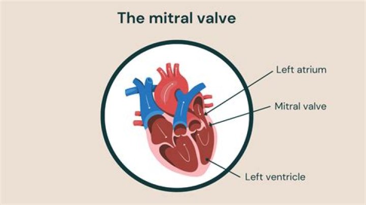
The mitral valve (/ˈmaɪtrəl/), also known as the bicuspid valve or left atrioventricular valve, is one of the four heart valves. It has two cusps or flaps and lies between the left atrium and the left ventricle of the heart.
What is mitral valve E A ratio?
The E/A ratio is the ratio of the early (E) to late (A) ventricular filling velocities. … The E/A ratio is measured by placing a pulsed wave Doppler across the mitral valve and measuring the velocities across the valve.
What is PISA method?
PISA Method Proximal Isovelocity Surface Area (PISA) is a method based off of flow convergence. If you missed our prior blog over flow convergence, you can find it here. This method is based off of the conservation of mass: flow through the regurgitant orifice = flow through the isovelocity surface.
How do you find the cross sectional area of an Lvot?
Answer: An LVOT diameter of 2 cm gives a LVOT cross-sectional area of, 2 * 2 * 0.78540 = 3.14 cm2. To calculate stroke volume, multiply the cross-sectional area of 3.14 cm2 by the LVOT VTI 24 cm.
What is a normal EE ratio?
In normal individuals the E/e´ ratio is <8. In the presence of diastolic dysfunction / impaired relaxation, e´ will be rather low. In contrast, the E-wave increases with elevated filling pressures. Thus the E/e´ ratio will increase in the presence of diastolic dysfunction.
What is AV Vmax?
Mild AS was defined as aortic valve (AV) thickening accompanied by a peak aortic jet velocity (AV Vmax) ≥2.0 and <3.0 m/sec, and rapid progression of AS was defined as an average annual increase in the AV Vmax ≥0.2 m/sec, and cardiac events were defined as cardiac death or AV replacement.
What is DVI in Echo?
The Doppler Velocity Index (DVI) is useful for assessing aortic prosthetic valve function as well as screening for valve obstruction. It is calculated as the ratio of the subvalvular velocity obtained by PW Doppler and the maximum velocity obtained by CW Doppler across the prosthetic valve.
How do you calculate CV for valves?
Cv by definition is the number of gallons per minute (GPM) a valve will flow with a 1 psi pressure drop across the valve. For example a valve with a Cv of 10 will flow 10 GPM with a 1 psi pressure drop. The formula used to select the valve Cv with the specified differential pressure is: Cv=GPM/((SQ RT(∆P)).
How is CV calculated?
The formula for the coefficient of variation is: Coefficient of Variation = (Standard Deviation / Mean) * 100. In symbols: CV = (SD/x̄) * 100. Multiplying the coefficient by 100 is an optional step to get a percentage, as opposed to a decimal.
What is CV and KV in control valve?
The Valve Coefficient (Cv – in Imperial unit) – the number of US GALLON PER MINUTE of water at 60 °F that will flow through a valve at specific opening with a pressure drop of 1 psi across the valve. The flow factor (Kv – in Metric unit) – the amount of water that will flow in m3/hr.
What is the normal aortic valve area?
In adult individuals with normal aortic valves, the valve area is 3.0 to 4.0 cm2. As aortic stenosis develops, minimal valve gradient is present until the orifice area becomes less than half of normal.
How do you calculate Mr regurgitant fraction?
Mitral regurgitant fraction (RF) was calculated as either RF(VOL) = [LVSV – RVSV] or RF(FLOW) = [LVSV – aortic flow volume], both expressed as a fraction of LVSV. The agreement of the measurements was assessed as a measure of robustness in clinical practice.
What is deceleration time of the mitral valve?
The flow profile of the mitral valve will have a Vmax, typically the maximum velocity of the E wave. The time from the Vmax to the where the velocity is equal to zero is the deceleration time.
What is the normal tricuspid valve area?
The valve consists of three leaflets, named after their positions: anterior, posterior and septal [Figure 2]. The normal valve area in adults is 4-6 cm2.
Why mitral valve is bicuspid?
… opening is guarded by the mitral, or bicuspid, valve, so named because it consists of two flaps. The mitral valve is attached in the same manner as the tricuspid, but it is stronger and thicker because the left ventricle is by nature a more powerful pump working under high pressure.
How many mitral valves are there?
The mitral valve is one of four valves in the heart that keep blood flowing in the right direction.
Why does mitral valve have 2 flaps?
Mitral Valve The leaflets open to let blood move forward through the heart during half of the heartbeat. They close to keep blood from flowing backward during the other half of the heartbeat. The mitral valve has only two leaflets; the aortic, pulmonic and tricuspid valves have three.
How is EE ratio calculated?
E/e′ ratio was calculated as E wave divided by e′ velocities. LV mass was estimated from LV linear dimensions and indexed to body surface area as recommended by ASE guidelines. LV hypertrophy was defined as LV mass indexed to body surface area (LV mass index) >115 g/m2 in men or >95 g/m2 in women.
What is Grade 3 left ventricular diastolic dysfunction?
Grade III – This is a severe form of diastolic dysfunction characterized by restrictive filling of the heart that leads to symptoms of advanced heart failure. When the patient is asked to perform the Valsalva manoeuvre during echocardiography, the diastolic abnormalities seem to reverse.
How do you measure severity of mitral regurgitation?
- Doppler color flow mapping is widely used in clinical practice to assess the severity of mitral regurgitation (MR), usually by the area of the jet projecting into the left atrium. …
- Regurgitant orifice area has been proposed as a marker of lesion severity in valvular regurgitation.