
What are contiguous leads in ECG
By Emily Dawson
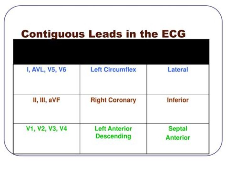
When we talk about reading a 12 lead EKG, we have to talk about the term contiguous. Contiguous leads are next to each other, anatomically speaking. They are all touching, and in the same general region (like the left ventricle, for example).
Which pair of leads would be considered contiguous leads?
Leads II, III and aVF are leads that have their positive electrode located at the left foot. They are contiguous leads that all look at the inferior wall of the left ventricle. Leads I and aVL are leads that have their positive electrode located on the left arm.
What is contiguous lead?
Contiguous leads are next to each other, anatomically speaking. They are all touching, and in the same general region (like the left ventricle, for example).
What are the 3 ECG leads?
For a routine analysis of the heart’s electrical activity an ECG recorded from 12 separate leads is used. A 12-lead ECG consists of three bipolar limb leads (I, II, and III), the unipolar limb leads (AVR, AVL, and AVF), and six unipolar chest leads, also called precordial or V leads, ( , , , , , and ).What is aVR lead?
In pericarditis, lead aVR is most often the only lead which shows reciprocal ST depression where as in Acute Infarction, usually a group of leads shows reciprocal depression. In the presence of persistent ST elevation in anterior chest leads, the R in aVR is suggestive of left ventricular aneurysm (Goldburger’s sign).
What are the anterior leads?
The arrangement of the leads produces the following anatomical relationships: leads II, III, and aVF view the inferior surface of the heart; leads V1 to V4 view the anterior surface; leads I, aVL, V5, and V6 view the lateral surface; and leads V1 and aVR look through the right atrium directly into the cavity of the …
What does V1 V2 V3 mean in ECG?
The areas represented on the ECG are summarized below: V1, V2 = RV. V3, V4 = septum. V5, V6 = L side of the heart. Lead I = L side of the heart.
How is MI diagnosed on ECG?
- ST segment elevation in the anterior leads (V3 and V4) at the J point and sometimes in the septal or lateral leads, depending on the extent of the MI. …
- Reciprocal ST segment depression in the inferior leads (II, III and aVF).
Why it is called 12 lead ECG?
In other words, each ECG lead is computed by analysing the electrical currents detected by several electrodes. The standard ECG – which is referred to as a 12-lead ECG since it includes 12 leads – is obtained using 10 electrodes.
Why do we use lead 2 in ECG?The most commonly used lead is lead II – a bipolar lead with electrodes on the right arm and left leg. This is the most useful lead for detecting cardiac arrhythmias as it lies close to the cardiac axis (the overall direction of electrical movement) and allows the best view of P and R waves.
Article first time published onHow is STEMI diagnosed?
Classically, STEMI is diagnosed if there is >1-2mm of ST elevation in two contiguous leads on the ECG or new LBBB with a clinical picture consistent with ischemic chest pain. Classically the ST elevations are described as “tombstone” and concave or “upwards” in appearance.
What are bipolar leads?
[ lēd ] n. The electrical connection of two electrodes to a recording instrument and to two different places on the body, such as the chest and a limb. A record obtained from the combined input of the two electrodes.
What is unipolar lead?
[ lēd ] n. A lead of an electrocardiograph in which one electrode is placed on the chest in the vicinity of the heart or on one of the limbs, while the other is placed at an area of zero potential. A record obtained from such a lead.
What are unipolar leads in ECG?
Unipolar leads (augmented leads and chest leads) have a single positive recording electrode and utilize a combination of the other electrodes to serve as a composite negative electrode. Normally, when an ECG is recorded, all leads are recorded simultaneously, giving rise to what is called a 12-lead ECG.
Where does V5 lead go?
V5 is placed directly between V4 and V6. V6 is placed over the fifth intercostal space at the mid-axillary line (as if drawing a line down from the armpit). V4-V6 should line up horizontally along the fifth intercostal space.
Which ECG machine is best?
ProductPriceProduct BPL 9108D 12 Channel ECG MachinePrice ₹93,000.00Product BPL Cardiart 9108,12 channel ECG MachinePrice ₹99,500.00Product Philips TC20 12 channel ECG MachinePrice ₹158,000.00Product 12 channel ECG Machine , Bionet Model Cardiocare 2000Price ₹60,200.00
What does it mean if you have an inverted T wave?
Despite this fact, inverted T waves in the setting of an appropriate clinical history are very suggestive of ischemia. Ischemia can be due to an acute coronary syndrome caused by rupture of an atherosclerotic plaque or due to factors increasing oxygen demand or decreasing oxygen supply such as severe anemia or sepsis.
Can you wear a bra during an ECG?
When you go for an ECG test, you will need to remove your upper clothing so that electrodes can be attached to your chest and limbs., Wearing a separate top with trousers or a skirt can allow easy access to the chest. Underwire in a bra can interfere with the ECG reading – you may be asked to remove it before the test.
What counts as ST elevation?
An ST elevation is considered significant if the vertical distance inside the ECG trace and the baseline at a point 0.04 seconds after the J-point is at least 0.1 mV (usually representing 1 mm or 1 small square) in a limb lead or 0.2 mV (2 mm or 2 small squares) in a precordial lead.
What are the 5 types of myocardial infarction?
ST segment elevation myocardial infarction (STEMI) non-ST segment elevation myocardial infarction (NSTEMI) coronary spasm, or unstable angina.
What are the causes of MI?
- Coronary occlusion secondary to vasculitis.
- Ventricular hypertrophy (eg, left ventricular hypertrophy, hypertrophic cardiomyopathy)
- Coronary artery emboli, secondary to cholesterol, air, or the products of sepsis.
- Coronary trauma.
Why does Mi cause ST elevation?
Accordingly, ST segment elevation during acute myocardial infarction requires the injury current to flow in the opposite direction [12, 13, 24], which can be caused by greater depression of the epicardial action potential.
What is EASI lead placement?
EASI lead placement: E, electrode placed on the lower part of the sternum, level with the fifth intercostal space; A, electrode placed at the level of the fifth intercostal space, on the left midaxillary line; S, electrode placed on the upper part of the sternum; I, electrode placed at the fifth intercostal space, on …
When is a 3-lead ECG used?
3-lead ECGs are used most often for recording a 24-hour reading. A 24-hour reading is a frequently used tool for the diagnosis of heart problems and is reimbursed as a long-term reading.
What is the difference between a 3 and 5 lead ECG?
5-lead monitoring is the same as 3-lead monitoring, but with two additional electrodes that enable the monitoring of extra leads and help improve ST elevation readings (Cables and Sensors 2016).
What is PCI medical?
Percutaneous coronary intervention (PCI) refers to a family of minimally invasive procedures used to open clogged coronary arteries (those that deliver blood to the heart). By restoring blood flow, the treatment can improve symptoms of blocked arteries, such as chest pain or shortness of breath.
What are the 3 cardiac enzymes?
Cardiac enzymes ― also known as cardiac biomarkers ― include myoglobin, troponin and creatine kinase.
Can STEMI present without chest pain?
As in patients with chest pain, there was a clear gradation of risk that was determined by the severity of the manifestation of the ACS, with almost 20% of STEMI patients presenting without chest pain dying in the hospital.
What are the 3 bipolar leads?
The bipolar extremity leads are called I, II and III. The unipolar extremity leads are called avR, avL and avF, and the chest leads are called V1–V6.
What is the difference between unipolar and bipolar leads?
A unipolar lead is a single conductor lead with an electrode located at the tip. A bipolar lead has two separate and isolated conductors within a single-lead; the distal electrode is located at the tip of the lead and the other one is usually about 2 cm more proximal.
What is augmented lead?
Augmented limb leads. The potential difference between the reference electrode and an exploring electrode constitutes the augmented lead. The three exploring electrodes are the right arm (R), the left arm (L), and the left leg (F).