
What are the 3 layers of tissue that form the eyeball
By Sophia Carter
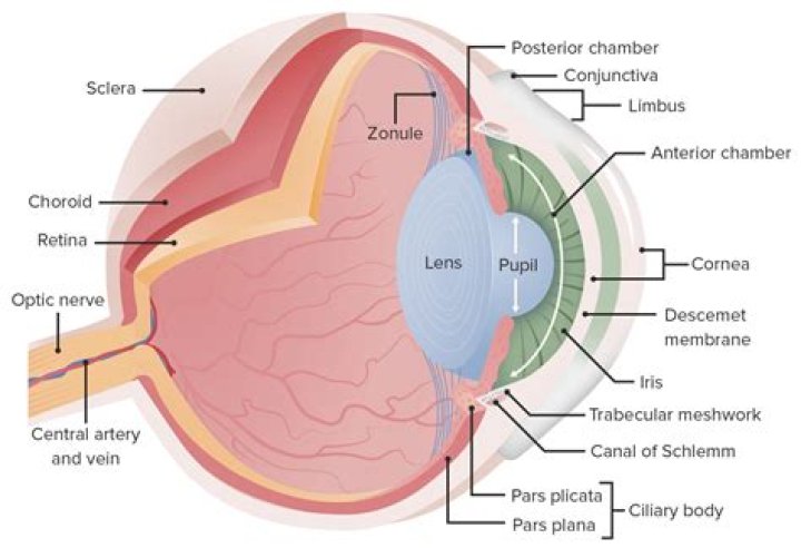
The outer layer of the eyeball is a tough, white, opaque membrane called the sclera (the white of the eye). … The middle layer is the choroid. … The inner layer is the retina, which lines the back two-thirds of the eyeball.
What are the 3 tissue layers of the eye?
- The sclera and cornea make up the exterior layers.
- The uvea is the vascular layer in the middle, subdivided into the iris, ciliary body, and choroid.
- The retina constitutes the innermost layer and is made up of nervous tissue.
Is eyeball a tissue?
The eye is a fluid-filled sphere enclosed by three layers of tissue (Figure 11.1). Most of the outer layer is composed of a tough white fibrous tissue, the sclera.
What tissue is the eyeball made of?
The eye’s outer layer is made of dense connective tissue, which protects the eyeball and maintains its shape. It is also known as the fibrous tunic. The fibrous tunic is composed of the sclera and the cornea. The sclera covers nearly the entire surface of the eyeball.What are the three layers of the eye quizlet?
What are the three layers of the eye? The sclera, the choroid layer, and the retina.
What is the parts of the eye?
Articles On Eye Basics Cornea: a clear dome over the iris. Pupil: the black circular opening in the iris that lets light in. Sclera: the white of your eye. Conjunctiva: a thin layer of tissue that covers the entire front of your eye, except for the cornea.
What are the layers of the eye and their functions?
The eye is made up of three layers: the outer layer called the fibrous tunic, which consists of the sclera and the cornea; the middle layer responsible for nourishment, called the vascular tunic, which consists of the iris, the choroid, and the ciliary body; and the inner layer of photoreceptors and neurons called the …
What is the outer layer of the eye quizlet?
The outermost tissue layer of the eye includes the sclera, and the cornea. The part of the fibrous tunic that is embedded in the eye socket is a thick, opaque, white layer called the sclera.What are eyeballs?
eyeball, spheroidal structure containing sense receptors for vision, found in all vertebrates and constructed much like a simple camera. … Much of the eyeball is filled with a transparent gel-like material, called the vitreous humour, that helps to maintain the spheroidal shape.
What layer of the eye is superficial and avascular?Cornea and sclera constitute the outer covering or coat of the eyeball. The main purpose of this coat is to protect structures inside the eye. The cornea is a transparent avascular tissue that acts as a structural barrier and protects the eye against infections.
Article first time published onWhat are the layers of the eye in order from superficial to deep?
From deep to superficial, they are the inner limiting membrane, nerve fiber layer, ganglion cell layer, inner plexiform layer, inner nuclear layer, outer plexiform layer, outer nuclear layer, external limiting membrane, and the retinal pigment epithelium.
Which structures are part of the vascular layer of the eyeball?
The vascular layer of the eye lies underneath the fibrous layer. It consists of the choroid, ciliary body and iris: Choroid – layer of connective tissue and blood vessels. It provides nourishment to the outer layers of the retina.
What are the three layers of the retina what takes place in each layer?
The cellular layers of the retina are as follows: 1) The pigmented epithelium, which is adjacent to the choroid, absorbs light to reduce back reflection of light onto the retina, 2) the photoreceptor layer contains photosensitive outer segments of rods and cones, 3) the outer nuclear layer contains cell bodies of the …
What is the innermost layer and most delicate layer of the eyeball where photoreceptors are located?
The retina is the innermost layer in the eye that is responsible for the visual processing that turns light energy from photons into three-dimensional images.
Which layer of the eye contains rods and cones?
Retina: a light sensitive layer that lines the interior of the eye. It is composed of light sensitive cells known as rods and cones.
How many parts are in the human eye?
The eye itself is made of 10 general components that all work together to keep us seeing well every day.
Where are rods and cones located?
The retina of the eye has two types of light-sensitive cells called rods and cones, both found in layer at the back of your eye which processes images.
What is the eyeball Emoji?
👀 Eyes emoji It mostly serves to draw attention to something the user wants to highlight, especially in situations that involve drama and interpersonal tension. It can also be an emoji representation of shifty eyes or the action of side-eyeing. This emoji sometimes appears when someone finds a person attractive.
What is the inner layer of the eye and contains the nerve cells rods and cones and bipolar cells?
The components of the neural retina. The neural retina consists of at least five different types of neurons: the photoreceptors (rods and cones), horizontal cell, bipolar cell, amacrine cell and ganglion cell.
What is ciliary epithelium?
The ciliary body is a part of the eye that includes the ciliary muscle, which controls the shape of the lens, and the ciliary epithelium, which produces the aqueous humor. The aqueous humor is produced in the non-pigmented portion of the ciliary body.
How many layers does the conjunctiva have?
ConjunctivaNumber of layersCells in the layersLimbal10 layers of stratified squamous epitheliumBasal- cubical
Which order is the correct pathway of vision?
The visual pathway consists of the retina, optic nerves, optic chiasm, optic tracts, lateral geniculate bodies, optic radiations, and visual cortex.
What is the outermost layer of the retina?
The center of the macula is called the fovea. The inner surface of the retina is adjacent to the vitreous of the eye. The outermost layer of the retina, the retinal pigment epithelium, is tightly attached to the choroid.
What is the vascular layer?
The vascular layer of the eye lying between the retina and sclera. This layer furnishes nourishment to outer layers of the retina.
Which structures are part of the outer fibrous layer of the eyeball quizlet?
- fibrous layer. the outermost layer of the eyeball, composed of the sclera and the cornea.
- sclera. …
- cornea. …
- vascular layer. …
- choroid. …
- ciliary body. …
- ciliary muscles. …
- ciliary processes.
Is the pupil in the vascular layer?
The vascular tunic of the eye is formed from behind forward by the choroid, the ciliary body, and the iris. … The iris is a circular diaphragm behind the cornea, and presents near its center a rounded aperture, the pupil.
What are the neural layers of the retina?
The neural retina consists of the inner limiting membrane (ILM), nerve fiber layer (NFL), ganglion cell layer, inner plexiform layer (IPL; synaptic processes between bipolar cells and ganglion cells), inner nuclear layer (INL; nuclei of bipolar, horizontal, amacrine, and Muller cells), outer plexiform layer (OPL; …
What is the correct order of the three retina cell layers starting with the layer that is closest to the iris?
Photoreceptor cells, ganglion cells, bipolar cells.
Which layer of the eyeball is the most delicate?
This is because the lens becomes less elastic with age. The retina (the soft, light-sensitive layer of tissue that lines the back of the eyeball wall) is made up of millions of light receptors called rods and cones. Rods are much more sensitive to light than cones.
What does the choroid layer do?
The choroid supplies the outer retina with nutrients, and maintains the temperature and volume of the eye. The choroidal circulation, which accounts for 85% of the total blood flow in the eye, is a high-flow system with relatively low oxygen content.
What is the name of the innermost membrane present in the human eye sclera choroid retina blindspot?
NEET Related LinksNEET Physics SyllabusNEET 2021 Updates