
What is a retinal Operculum
By Olivia House
The operculum, which can be seen as a whitish disc-shaped floater, remains attached to the posterior hyaloid membrane overlying the retinal hole. With time, the operculum contracts and deteriorates because of loss of retinal blood supply. In many cases, it is observed to be smaller than the underlying retinal defect.
What causes a retinal tuft?
They are caused by vitreous opacities, such as epipapillary glial tissue torn from the optic disc, condensations of vitreous collagen, and/or blood. There are no symptoms that can distinguish a PVD alone from a PVD with an associated retinal break.
How are retinal holes treated?
Vitrectomy is the most common treatment for macular holes. In this surgical procedure, the vitreous gel is removed to stop it from pulling on the retina, and most commonly a gas bubble is placed in the eye to gently hold the edges of the macular hole closed until it heals.
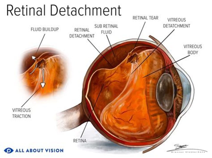
How serious is a retinal hole?
Retinal holes and tears do not automatically cause severe vision problems; instead, the areas of the retina that have holes and tears will not function correctly. If the condition is not well managed or treated in due time, then significant vision loss or even blindness can occur.How common is detached retina after cataract surgery?
Among the advanced procedures we provide are surgeries to treat retinal detachment. Although relatively rare, retinal detachment is a very real risk of cataract surgery, occurring in roughly 1 percent of post-operative cataract surgery patients.
Can retinal holes heal themselves?
Although some macular holes heal on their own without treatment, in many cases, surgery is necessary to improve vision. The surgery eye doctors use to treat this condition is called a vitrectomy. During a vitrectomy, the vitreous gel is removed to prevent it from pulling on the retina.
How common are retinal Tufts?
Cystic Retinal Tufts These are noted in approximately 5% of the population and are thought to be a congenital abnormality in the development of the peripheral retina.
How long does it take to heal from a vitrectomy?
You might have some pain in your eye and your vision may be blurry for a few days after the surgery. You will need 2 to 4 weeks to recover before you can do your normal activities again. It may take longer for your vision to get back to normal.Is a retinal hole an emergency?
Certain changes in your vision can indicate serious damage to the retina, such as holes and tears or detached retina, an emergency situation in which you can permanently lose your vision. Retinal detachment itself is painless, but there are almost always warning signs.
How long do I have to stay face down after vitrectomy?Patients having vitreo-retinal surgery for a macular hole will need to posture face down for 14 days; for other conditions this is only necessary for 5 days.
Article first time published onIs retinal laser surgery painful?
Pain: Most patients have little if any pain following retinal laser surgery. Patients who require more extensive laser may have an ache inside the eye or around the eye. If you have discomfort after the surgery, rest and take Tylenol, ibuprofen, or another over the counter pain reliever.
What happens if you don't stay face down after vitrectomy?
For example, facedown positioning has the potential to cause mesenteric venous obstructions. Additionally, patients who are hypercoagulable can develop deep vein thrombosis or pulmonary embolism.
What is the success rate of retinal detachment surgery?
In most specialist centres around nine out of ten retinal detachments are successfully repaired with a single operation. In the remaining cases, the retina re-detaches and needs another operation. The final success rate is over 95 per cent.
How do you fix retinal detachment?
One method of retinal detachment repair is pneumatic retinopexy. In this procedure, a gas bubble is injected into the eye. The bubble presses against the detached retina and pushes it back into place. A laser or cryotherapy is then used to reattach the retina firmly into place.
Does cataract surgery weaken the retina?
The retina is located in the interior of the back of the eye, and cataract surgery does not interfere with this area. The surgery, most doctors maintain, will not improve vision lost from retinal degeneration, but it will not make the retinal condition worse.
Can the retina be damaged during cataract surgery?
Cataract surgery, like any surgical procedure, has associated complications. Acute retinal complications include globe perforation, dislocated lens fragments, hemorrhagic choroidal detachment, and endophthalmitis.
What are the side effects of barrage laser treatment?
Complications of laser barrage include transient effects (blurred vision, raised intraocular pressure, and headache) and permanent effects (poor night vision, poor color vision, and peripheral field defect).
What is retinal dialysis?
A retinal dialysis is a tear in the retina whose anterior edge is at the ora serrata and whose posterior edge is attached to the vitreous base. 1. The majority of patients presenting for surgical management of retinal dialysis are known to have a significant past history of trauma to the affected eye.
What are retinal Tufts?
A tuft on the retina is a small elevation where the bag of gel that fills the eye is pulling on a focal part of the retina. However there is no tear in the retina.
Will macular hole get worse?
If you do have a macular hole and you don’t seek help, your central vision will probably get gradually worse. Relatively early treatment (within months) may give a better outcome in terms of improvement in vision.
What does your vision look like with a retinal tear?
A sudden appearance of light flashes, which could be the first stage of a retinal tear or detachment. Having a shadow appear in your peripheral (side) field of vision. Seeing a gray curtain slowly moving across your field of vision. Experiencing a sudden decrease in vision, including focusing trouble and blurred vision.
Can an optometrist see a retinal tear?
These routine vision tests do not detect retinal detachment, but they can find problems that could lead to or result from retinal detachment. A doctor can usually see a retinal tear or detachment while examining the retina using ophthalmoscopy.
Is retinal detachment surgery painful?
Surgery is done under anesthesia, so it’s not painful. After surgery, you may have some amount of pain in the eye. Your eye may be tender, red or swollen for a couple of weeks.
Is a vitrectomy a serious operation?
Vitrectomy procedures are an effective surgery and severe complications are rare. According to the American Society of Retina Specialists, most surgeries have a 90 percent success rate.
How serious is a vitrectomy?
If not treated, some of them can even result in blindness. In some cases, vitrectomy can restore lost vision. You might need a vitrectomy done in an emergency — an eye injury, for example.
Does vitreous come back after vitrectomy?
The vitreous humor cannot regenerate; therefore, the cavity must be filled with a substitute material during and after vitrectomy. Natural polymers, although a reasonable choice for a vitreous substitute, are limited by low stability.
Is a vitrectomy painful?
Unless the patient is in poor health or has severe disease, nearly all vitrectomies are outpatient procedures performed either in a hospital or in a dedicated ambulatory surgery center; they involve little or no pain and require only minimal anesthesia.
Do you have to sleep face down after vitrectomy?
Face down (“eyes down”) posturing is only required during waking hours, not when you’re sleeping. It is recommended to sleep on either side or even your front, but not sleep on your back as that would make the bubble move away from the macular hole.
How much does a vitrectomy cost?
How Much Does a Vitrectomy Cost? On MDsave, the cost of a Vitrectomy ranges from $7,603 to $9,232. Those on high deductible health plans or without insurance can save when they buy their procedure upfront through MDsave.
How long does it take to regain vision after retinal surgery?
In fact complete healing after retinal surgery often takes 6 months. In most cases, the visual acuity at 6 months will be the final vision. There is normal swelling of the eye after retina surgery, which initially, will limit the vision.
Is it normal to see black dots after retinal surgery?
With laser treatment, black dots are common. Expect your eye to be swollen. If you are posturing after surgery often this swelling can get worse after the first day and can affect the fellow eye. This is quite normal and can look like one large “blister” on your eye lid.