What is the atrial syncytium
By Sophia Carter
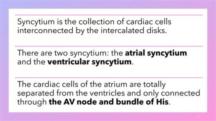
Definition. A network of cardiac muscle cells connected by gap junctions that allows coordinate contraction of the atria. Supplement. There are two syncytia of the heart: the ventricular syncytium and the atrial syncytium separated by fibrous tissue.
What are the atrial syncytium and ventricular syncytium?
Muscles of the atria and those of the ventricles are arranged to form an atrial and ventricular syncytium. syncytium is an arrangement of muscle fibers in which the fibers fuse to form an interconnected mass of fibers.
Why is heart called functional syncytium?
The mass is the result of surrounding cells fusing together into one larger cell. In cardiac tissue, the individual muscle cells do not fuse this way. Instead, they are linked together to form a mass of tissue that functions as if it were one large cell, which is why it is called a functional syncytium.
What is the function of syncytium?
The syncytium of cardiac muscle is important because it allows rapid coordinated contraction of muscles along their entire length. Cardiac action potentials propagate along the surface of the muscle fiber from the point of synaptic contact through intercalated discs.How many syncytium are there in a human heart?
The heart is composed of two separate functional syncytiums, the atrial syncytium and the ventricular syncytium.
What characteristic of a muscle cell is described as a syncytium?
The Skeletal Muscle Fiber. Skeletal muscle cells or fibers are highly elongated cells with a very elastic and resistant plasma membrane, called the sarcolemma. Fibers are characterized by the presence of numerous nuclei located at the periphery of the cell, hence muscle fibers are described as a syncytium.
What is syncytial arrangement?
A large cell-like structure formed by the joining together of two or more cells. The plural is syncytia.
What is an advantage of a syncytium?
Syncytia in Humans These fibers are individual, long syncytia. One advantage of this structure is fast communication and response between the brain and the muscles. The lack of separate membranes allows the impulses from the brain to quickly move between nuclei.What happens syncytium?
The syncytium As first visible changes in the ISC callose-like material is deposited along the cell wall where the stylet is inserted [2], cytoplasmic streaming is accelerated, and the nucleus is enlarged. In further changes most organelles are involved.
What is the meaning of Coenocyte?Definition of coenocyte 1a : a multinucleate mass of protoplasm resulting from repeated nuclear division unaccompanied by cell fission. b : an organism consisting of such a structure. 2 : syncytium sense 1.
Article first time published onIs heart a functional syncytium?
Cardiac muscle behaves as a functional syncytium, although it is composed of individual cells. At the lateral regions of the intercalated disks, gap junctions are protected from forces during contraction.
Where is the atrial syncytium?
A network of cardiac muscle cells connected by gap junctions that allows coordinate contraction of the atria. There are two syncytia of the heart: the ventricular syncytium and the atrial syncytium separated by fibrous tissue.
What is the function of myocardium?
Cardiac muscle tissue, or myocardium, is a specialized type of muscle tissue that forms the heart. This muscle tissue, which contracts and releases involuntarily, is responsible for keeping the heart pumping blood around the body.
What is the difference between Coenocytic and syncytium?
A coenocyte (English: /ˈsiːnəsaɪt/) is a multinucleate cell which can result from multiple nuclear divisions without their accompanying cytokinesis, in contrast to a syncytium, which results from cellular aggregation followed by dissolution of the cell membranes inside the mass.
How does the cardiac muscle work as a syncytium?
This joining is called electric coupling, and in cardiac muscle it allows the quick transmission of action potentials and the coordinated contraction of the entire heart. This network of electrically connected cardiac muscle cells creates a functional unit of contraction called a syncytium.
What is the Bengali meaning of syncytium?
বিভিন্ন নিউক্লিয়াস ধারণকারী সাইটোপ্লাজমে একটি ভর এবং একটি ঝিল্লি ঘিরা কিন্তু কোন অভ্যন্তরীণ সেল সীমানা (পেশী fibers হিসেবে
What cell junction is found in the heart muscle?
Cardiac muscle cells are equipped with three distinct types of intercellular junction–gap junctions, “spot” desmosomes, and “sheet” desmosomes (or fasciae adherentes)–located in a specialized portion of the plasma membrane, the intercalated disk.
Which type of junction enables the smooth muscles to contract as a Syncytium?
Respiratory system: Airway smooth muscles, such as tracheal smooth muscle and bronchial smooth muscle, are known to form electrical syncytia by means of gap junctions. The gap junctions are reported to play an important role in the regulation of contractions in airway smooth muscle in response to physiological stimuli.
What cells are in skeletal muscle tissue?
Skeletal muscle cells are long, cylindrical, and striated. They are multi-nucleated meaning that they have more than one nucleus. This is because they are formed from the fusion of embryonic myoblasts. Each nucleus regulates the metabolic requirements of the sarcoplasm around it.
What is heart tissue called?
Cardiac muscle tissue is one of the three types of muscle tissue in your body. The other two types are skeletal muscle tissue and smooth muscle tissue. Cardiac muscle tissue is only found in your heart, where it performs coordinated contractions that allow your heart to pump blood through your circulatory system.
What events lead to formation of Syncytium?
Syncytia is formed by fusion of an infected cells with neighboring cells leading to the formation of multi-nucleate enlarged cells. This event is induced by surface expression of viral fusion protein that are fusogenic directly at the host cell membrane.
Which muscles are immune to fatigue?
Cardiac or heart muscle immune fatigue due to the presence of a high number of mitochondria than skeletal muscle. The high number of mitochondria provides a steady supply of energy to the heart, therefore the heart does not stop due to low energy.
Why is muscle Fibre a Syncytium?
Complete answer: Skeletal muscles are also known as striated muscles and striated muscles. These muscles are innervated by branches from cranial and spinal nerves. Intercalated discs in these muscles are usually absent. They are multinucleated muscles and hence syncytial.
What is Multinucleated cytoplasm?
Multinucleate cells (multinucleated or polynuclear cells) are eukaryotic cells that have more than one nucleus per cell, i.e., multiple nuclei share one common cytoplasm. … For example, slime molds have a vegetative, multinucleate life stage called a plasmodium.
What is Coenocytic mycelium Class 11?
Hint: Coenocytic refers to a structure of an organism or an organism itself, that has multiple nuclei in a continuous protoplasmic mass, enclosed by a cell membrane or cell wall. This condition is generally found in the case of algae or fungi.
What is meant by an Aseptate coenocyte?
Coenocytic hyphae are nonseptate, also called aseptate, meaning they are one long cell that is not divided into compartments. … Coenocytic hyphae result from nuclear divisions within a cell without an accompanying division of the cytoplasm (cytokinesis).
Which type of tissue forms the skeleton of the heart?
The fibrous skeleton is made up of dense connective tissue. It is the fibrous skeleton that allows the controlled contraction of the heart, as it provides the connective tissue skeleton necessary. In certain animals (for example sheep), the central connective tissue of the heart contains a central bone.
Where is the AV bundle located?
In the normal heart, the AV node is located in the triangle of Koch. This triangular area is located on the septal wall of the right atrium, between the tricuspid valve, coronary sinus orifice, and tendon of Todaro, and marks the site of the AV node.
What is the bundle of His and Purkinje Fibres?
Bundle of His is a collection of specialized heart muscle cells that transmit electrical impulses from the AV node in the heart to the muscle cells of the heart wall. Meanwhile, Purkinje fibres are thin filaments that distribute electrical impulses to the ventricle myocardium and activate right and left ventricles.
What is myocardium layer?
The muscles of the heart, termed the myocardium, make up the middle and thickest layer of the heart wall. This layer lies between the single-cell endocardium layer, which lines the inner chambers, and the outer epicardium, which makes up part of the pericardium that surrounds and protects the heart.
What is the function of the papillary muscles?
Background— The papillary muscles (PMs) play an important role in normal cardiac function, helping to prevent leakage through the AV valves during systole. The nature of their attachment to the heart wall can affect the understanding of their function.