What tendon attaches to the calcaneus
By Jessica Hardy
The Achilles tendon attaches to the calcaneal tubercle. The extensor digitorum brevis: It originates on the dorsolateral side of the calcaneus and provides extension of the second to fourth digits.
What attached to the calcaneus?
Three muscles insert on the calcaneus: the gastrocnemius, soleus, and plantaris. These muscles are part of the posterior compartment of the leg and aid in walking, running and jumping.
What tendon connects to the heel?
The Achilles tendon connects your calf muscles to your heel bone.
What ligaments are attached to the calcaneus?
Cervical ligament. Talocalcaneal interosseous ligament, Lateral, intermediate, and medial roots of the inferior extensor retinaculum. Bifurcate ligament.Does the Achilles tendon attach to the calcaneus?
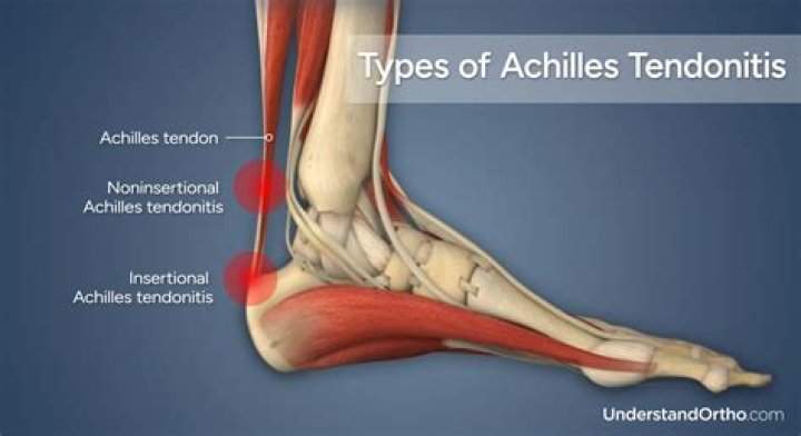
The Achilles (uh-KILL-ease) tendon is a band of tissue in the back of your leg. This tendon links your heel bone (calcaneus, pronounced cal-KAY-nee-us) to your calf muscles. It’s also called the calcaneal tendon.
Does your Achilles tendon heel?
When the calf muscles flex, the Achilles tendon pulls on the heel. This movement allows us to stand on our toes when walking, running, or jumping. Despite its strength, the Achilles tendon is also vulnerable to injury, due to its limited blood supply and the high tensions placed on it.
Which muscle attaches to the femur and calcaneus?
Gastrocnemius: The gastrocnemius, a two-headed muscle, is the most superficial of the muscles in the posterior compartment. Attachments: Both heads originate from the femur. The fibers converge to form the calcaneal tendon which attaches to the heel.
What is plantar anatomy?
45171. Anatomical terminology. The plantar fascia is the thick connective tissue (aponeurosis) which supports the arch on the bottom (plantar side) of the foot. It runs from the tuberosity of the calcaneus (heel bone) forward to the heads of the metatarsal bones (the bone between each toe and the bones of the mid-foot) …What inserts on the sustentaculum tali?
Several ligamentous structures attach to the sustentaculum tali: plantar calcaneonavicular ligament (anterior surface) deltoid ligament (medial surface) medial talocalcaneal ligament.
What is calcaneal bursitis?Calcaneal Bursitis (Subcutaneous Calcaneal Bursitis) Calcaneal bursitis is almost exclusively associated with shoes that dig into the back of the heel. This condition can cause a hard, red bump to develop at the back of the heel.
Article first time published onWhat muscle is anterior to the Achilles calcaneal tendon?
Achilles tendonThe Achilles tendon or calcaneal tendon is attached to the gastrocnemius and soleus muscles.DetailsLocationBack of the lower legIdentifiers
How do I know if I hurt my Achilles tendon?
- Pain down the back of your leg or near your heel.
- Pain that gets worse when you’re active.
- A stiff, sore Achilles tendon when you first get up.
- Pain in the tendon the day after exercising.
- Swelling with pain that gets worse as you’re active during the day.
Where does Achilles tendon attach?
The Achilles tendon is a thick tendon located in the back of the leg. It connects the gastrocnemius and soleus muscles in the calf to an insertion point at the calcaneus (heel bone).
Where does the calcaneal tendon insertion?
Achilles tendon, also called calcaneal tendon, strong tendon at the back of the heel that connects the calf muscles to the heel. The tendon is formed from the gastrocnemius and soleus muscles (the calf muscles) and is inserted into the heel bone.
What is the plantaris tendon?
The plantaris muscle is a fine rope-like tendon running next to the larger Achilles Tendon. Its function is to work with the Achilles to flex the ankle and knee joint by extending from the outside (lateral) back of the femur (allowing you to stand on your toes or point your foot).
What attaches to calcaneal tuberosity?
The Achilles tendon attaches to the calcaneal tubercle.
Does the fibula articulate with the calcaneus?
The talus articulates superiorly with the distal tibia, the medial malleolus of the tibia, and the lateral malleolus of the fibula to form the ankle joint. The talus articulates inferiorly with the calcaneus bone. The sustentaculum tali of the calcaneus helps to support the talus.
What bone is above the calcaneus?
Talus: also called the ankle bone, sits above the heel bone (calcaneus) and makes up the lower part of the ankle joint by connecting the tibia and fibula with the foot. Cuboid: a cube-shaped bone that connects the foot to the ankle and helps provide stability to the foot.
Where is Achilles heel located?
It is found in the back of the leg, at the bottom half of the calf. The Achilles consists of cords of fibrous tissue connecting the calf muscles to the back of the heel bone. This musculoskeletal structure allows you to walk, run, and move. Common problems with the achilles tendon include tendonitis and tendon rupture.
Is it OK to walk with Achilles tendonitis?
In all individuals, Achilles tendinopathy can result in a limited ability to walk, climb stairs, or participate in recreational activities.
What happens if Achilles tendonitis goes untreated?
What happens if Achilles tendonitis goes untreated? If left untreated, the condition of Achilles tendinitis usually gets worse. You will likely begin to feel chronic pain and the tendon may get ruptured. The condition could become very serious and could lead to serious injury.
What tendon goes under sustentaculum tali?
After passing through the tarsal tunnel, the flexor hallucis longus tendon must curve around a bony landmark called the sustentaculum tali.
What is calcaneus Secundarius?
The calcaneus secundarius (CS) is an accessory ossicle of the anterior facet of the calcaneus and is usually asymptomatic. This accessory bone can be frequently mistaken for a fracture of the anterior process of the calcaneus.
What is the talar shelf?
On the medial side of the base of the calcaneal tuberosity is the sustentaculum tali (talar shelf), a shelf like process that overlaps the plantar aspect of talus and supports the deep digital flexor tendon. On the plantar side of the sustentaculum tali is the groove for tendon of flechissor digitorum lateralis.
Does the Achilles tendon attach to the plantar fascia?
The Achilles tendon also attaches to the plantar fascia. If the calf muscles that attach to the Achilles tendon are tight, the ankle becomes less flexible, and the plantar fascia also tightens. Frequently, the pain is noticed after getting up in the morning or after standing after long periods of sitting.
What tendons are involved with plantar fasciitis?
Tight Achilles tendons, which are the tendons attaching your calf muscles to your heels, may also result in plantar fascia pain. Simply wearing shoes with soft soles and poor arch support can also result in plantar fasciitis. Plantar fasciitis isn’t typically the result of heel spurs.
Is the plantar fascia a tendon?
The plantar fascia is the flat band of tissue (ligament) that connects your heel bone to your toes. It supports the arch of your foot. If you strain your plantar fascia, it gets weak, swollen, and irritated (inflamed).
What is Haglund's syndrome?
Haglund’s deformity is an abnormality of the bone and soft tissues in the foot. An enlargement of the bony section of the heel (where the Achilles tendon is inserted) triggers this condition. The soft tissue near the back of the heel can become irritated when the large, bony lump rubs against rigid shoes.
What is the difference between Achilles tendonitis and Achilles bursitis?
Tendonitis is a painful condition where the tendons become inflamed. Bursitis is when the small sacs of fluid around a joint (called bursa) become irritated and inflamed. Both conditions can present with swelling and discomfort around the affected joints.
What is retrocalcaneal spur?
Extra bone formation (exostosis) on the back (retro) of the heel bone (calcaneus) is called a retrocalcaneal exostosis. Some people will also call this a heel spur, different from a spur found on the bottom of the heel. The extra bone develops on the back of the heel bone and within the Achilles tendon.
What does a soleus strain feel like?
Grade-one muscle strains, such as those experienced in the soleus, can be classified as mild pain with activity, localized tenderness on palpation, mild spasm and swelling.